

Fortis Cunningham Road removes 25 Abdominal Fibroids Weighing 14.9 Kgs from a 37-Year-old Woman in an Rare Surgical Feat

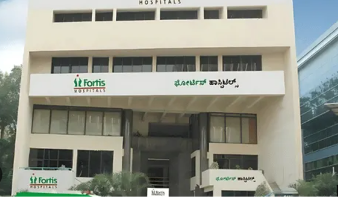
Bengaluru: Doctors at Fortis Hospital, Cunningham Road have successfully removed 25 fibroids with a combined weight of 14.9 kg, including a giant 6.1 kg mass from a 37-year-old woman. The patient was diagnosed with Disseminated Peritoneal Leiomyomatosis (DPL) – an exceptionally rare non-cancerous condition with fewer than 250 cases reported worldwide.
The patient had been delaying treatment for nearly six years due to fear of surgery, allowing the fibroids to grow extensively and severely distort her pelvic and abdominal anatomy. The highly complex surgery was performed by a team led by Dr. Rubina Shanawaz, Additional Director – Uro-Gynaecology, Gynaec-Oncology, Laparoscopic & Robotic Surgery, along with Dr. Punyashree R.M., Consultant – Gynaec-Oncology and Dr. Prakruthi S., Associate Consultant – Uro-Gynaecology.
The patient was presented at Fortis Hospital, Cunningham Road with severe abdominal swelling and discomfort. Given the rapid increase in the tumour mass and her previous surgical history, the doctors carried out detailed investigations, including MRI scans and blood tests, to assess the extent of the condition and rule out cancer. Detailed Investigations revealed a total of 25 fibroids across the abdomen, including a giant fibroid weighing up to 6.1 kg, which was extending up to the spleen. Further evaluations confirmed that the growths were non-cancerous fibroids.
The multi-disciplinary team of doctors successfully performed a highly complex surgery to remove the giant fibroid, along with several other fibroids spread across the abdomen. As the fibroids had significantly changed the normal position of the abdominal and pelvic organs, the procedure required meticulous planning and close coordination between the uro-gynaecology, anaesthesia and radiology teams. The doctors carefully removed the fibroids while preserving vital organs and critical structures. The patient recovered well, post-surgery and was discharged in a stable condition.
The case highlights the importance of timely evaluation and appropriate management of fibroids, particularly when symptoms are persistent or there is progressive abdominal enlargement. The extensive size and distribution of the fibroids, made the surgery particularly complex and required coordinated multi-disciplinary care.
Giving details of the case, Dr. Rubina Shanawaz, Additional Director, Uro-Gynaecology, Gynaec-Oncology, Laparoscopic & Robotic Surgery, Fortis Hospital Cunningham Road said, “Disseminated Peritoneal Leiomyomatosis is an extremely rare condition in which multiple fibroids form throughout the abdomen and can closely mimic advanced cancer. In this patient, the presence of 25 fibroids, including a giant 6.1-kg mass extending up to the spleen, had significantly distorted the normal abdominal and pelvic anatomy. The surgery therefore required meticulous dissection, careful preservation of vital structures and close coordination across specialties. Careful planning before surgery and testing during the procedure helped guide the surgery and ensure the safe removal of all the fibroids.”
Dr. Punyashree R.M, Consultant- Gynaec-Oncology, Fortis Hospital Cunningham Road said, “While fibroids are common and usually non-cancerous, their symptoms can vary significantly from one woman to another. Persistent heavy menstrual bleeding, pelvic discomfort, abdominal swelling or a noticeable increase in abdominal size should not be ignored, particularly when symptoms progressively worsen. Timely evaluation can help identify the condition early and allow doctors to determine the most appropriate treatment before the fibroids become significantly enlarged or begin affecting surrounding organs.”
Dr. Prakruthi S., Associate Consultant – Uro-Gynaecology, Fortis Hospital Cunningham Road, said, “In cases where fibroids are unusually large or present in multiple locations, a detailed evaluation is essential to understand their size, location and impact on surrounding organs. Imaging plays an important role in assessing the extent of the disease and planning the surgical approach. In rare cases such as this, distinguishing benign fibroid disease from conditions that may resemble cancer is particularly important, and a combination of clinical assessment, imaging and appropriate pathological evaluation helps us plan treatment safely and effectively.”
