

Persistent Cough Reveals Hidden Foreign Body: Minimally Invasive Thoracoscopy helps 10-Year-Old Girl Recover

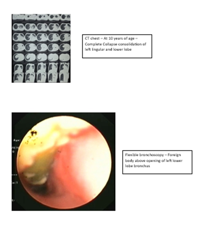
Mumbai: In a recent case involving a 10-year-old girl, a persistent cough since early childhood had resulted in a host of unanswered questions. Mysha had been receiving treatment for suspected tuberculosis, followed by asthma and pneumonias, but the cough did improve and kept recurring. When she came to Narayana Health SRCC Children’s Hospital, Mumbai, the team of doctors found that repeated scans had caused progressive damage to her left lung. They finally uncovered the real cause—a foreign body lodged deep inside the child’s airway for several years.
A plan was made to remove the deeply impacted forgotten foreign body of over 7 years through bronchoscopy. A multidisciplinary team led by Dr Indu Khosla, Senior Consultant, Paediatric Pulmonology, Narayana Health SRCC Children’s Hospital was set up which included the team of pulmonology, anaesthetists, and ENT surgeons. Following successful removal of the obstruction and a minimally invasive thoracoscopic lobectomy surgery, the 10-year-old is now breathing better and is on the road to recovery.
Mysha had an uneventful birth and healthy early childhood. However, at the age of three and a half years, she developed a persistent cough and fever, accompanied by poor appetite and weight loss. Chest X-rays and CT scans raised a clinical suspicion of tuberculosis, and she was started on treatment for TB. When her symptoms failed to improve, she was treated with inhalers for suspected asthma. Despite multiple treatments, she struggled with recurrent respiratory problems and poor weight gain.
Over the next five years, serial CT scans had revealed worsening damage to the same part of her lung, with collapse and consolidation of the left lower lobe and a healthy upper lobe expansion to compensate for the collapsed lower lobe. The gradual deterioration of the same lung, despite prolonged treatment, prompted the team of medical experts to investigate further.
After carefully reviewing her clinical history and imaging, doctors suspected a long-standing foreign body aspiration. A flexible bronchoscopy confirmed their suspicion, revealing a foreign body lodged just above the opening of the left lower lobe bronchus. The object was successfully removed using a combination of flexible and rigid bronchoscopy without any complications.
Although the obstruction had been cleared, years of blockage had already caused irreversible damage to the affected lung, leading to bronchiectasis (the airways become permanently wide). and damaged, thus prone to infections, and necrosis (localised death of lung tissue) of the left lower lobe.
To preserve the remaining healthy lung tissue and reduce the risk of recurrent infections, the multidisciplinary team performed a thoracoscopic left lower lobectomy. The damaged portion of the lung was successfully removed through a minimally invasive approach while preserving the healthy surrounding tissue. The surgery was uneventful, without any postoperative complications, and Mysha was discharged five days later.
Post-op, Mysha was started on airway clearance therapy to improve lung function. At her one-month follow-up, she showed encouraging clinical improvement, including better respiratory health and healthy weight gain.
Explaining the case, Dr Indu Khosla said, “Foreign body aspiration can sometimes remain undetected for years, particularly when the initial choking episode is not witnessed or remembered. In children with persistent cough, recurrent chest infections, or repeated collapse of the same part of the lung despite appropriate treatment, clinicians should maintain a high index of suspicion for an occult airway foreign body. Early diagnosis is essential to prevent irreversible lung damage, and when there is a suspicion, a flexible bronchoscopy check always helps. Even in delayed cases, timely bronchoscopic removal, appropriate pulmonary rehabilitation, and surgery when indicated can significantly improve long-term outcomes and quality of life.”
Sometimes, the most persistent symptoms have the most unexpected causes. This case highlights the importance of looking beyond common diagnoses when respiratory symptoms fail to improve as expected. It also demonstrates how careful clinical evaluation, advanced bronchoscopy, and minimally invasive surgery can together transform the outcome for a child, preserving healthy lung tissue and giving her the opportunity to breathe easier and lead a healthier life.
