

Rare Chest Wall Reconstruction Surgery for unprotected heart on 3-Month-Old Baby Performed Successfully

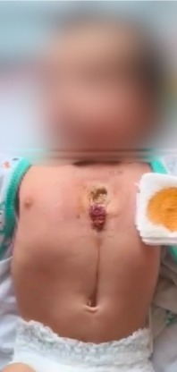
Mumbai : When Baby Janki was born, her parents immediately sensed something was unusual. Instead of the protective breastbone that normally shields the heart, there was a visible gap in the centre of her tiny chest. With every heartbeat, they could actually see her heart pulsating beneath the skin—a distressing sight for any parent and a reminder of how vulnerable their newborn was.
At just three months and twenty days old, Janki was diagnosed with a partial congenital sternal cleft, an exceptionally rare birth defect in which the breastbone fails to fuse during foetal development. Although she appeared active, fed well, and showed intermittent signs of striodor and breathlessness, her exposed heart remained at constant risk of injury. What seemed like a stable condition on the surface concealed a potentially life-threatening congenital anomaly that required timely surgical correction.
The condition had not been detected during pregnancy, making the diagnosis even more unexpected for the family. In the weeks following birth, Janki experienced only one episode of respiratory infection, which resolved with antibiotics. Apart from intermittent noisy breathing, she showed none of the symptoms that are often associated with complex congenital abnormalities. Yet the failure of fusion of the two halves of sternum meant that the heart and major blood vessels lacked their natural protection, leaving even minor trauma capable of causing devastating consequences.
When Janki was brought to Narayana Health SRCC Children’s Hospital, Mumbai, a multidisciplinary team immediately began a detailed evaluation to understand the extent of the defect and plan the safest course of treatment.
Clinical examination confirmed a partial midline sternal cleft, where two halves of superior part of the sternum where separated by nearly five centimetres. Despite the severity of the deformity, her oxygen levels remained normal, and her heart function appeared stable without any murmur. To better understand the anatomy, specialists performed a computed tomography pulmonary angiography (CTPA), which provided a detailed three-dimensional assessment of the chest.
The imaging confirmed the complete absence of the fused sternum and also revealed mild enlargement of the right side of the heart, along with small areas of lung collapse and patchy lung consolidation. Importantly, the major blood vessels and airways were otherwise normal. These findings enabled surgeons to carefully map out a reconstruction strategy tailored to the infant’s delicate anatomy.
Given the rarity and complexity of the condition, specialists Dr. Rasiklal Shah from Senior Consultant Paediatric Surgery, Dr. Pradeep Kaushik Senior consultant Paediatric Cardiothoracic and Congenital Heart Surgery, Senior Consultant Paediatric Cardiac Anesthesiology Dr. Nandini Dave and her team, Radiologists Dr. Hiren Panwala and Dr. Sonal Garg, and Paediatric Cardiac Intensive Care came together to design a comprehensive treatment plan. Every step was meticulously coordinated, with the shared goal of protecting the heart, restoring the chest wall, and ensuring the baby could grow and develop normally.
The infant underwent complex sternal reconstruction under general anaesthesia in a highly planned procedure. Recognising the potential risks associated with operating so close to the heart, the surgical team established advanced monitoring and kept a cardiopulmonary bypass machine on standby throughout the operation as an added safety measure.
During surgery, the chest wall was carefully reconstructed using a combination of advanced techniques. The surgeons mobilised the pectoralis major muscles to create protective tissue coverage, performed carefully planned chondrotomies of the clavicles and upper ribs to allow the chest to be brought together without excessive tension, and reconstructed the sternum using steel wires to restore the natural framework of the chest.
Although temporary fluctuations in blood pressure occurred while mobilising the chest structures—a known challenge during such intricate procedures—the experienced team of paediatric anaesthesiologists and paediatric cardiac teams managed the situation seamlessly, allowing the surgery to proceed safely.
Following the operation, Janki was closely monitored in the Paediatric Cardiac Intensive Care Unit for four days before being shifted to the ward. Her recovery progressed remarkably well. She resumed full oral feeds, remained haemodynamically stable throughout her hospital stay, and showed no postoperative complications. Eight days after surgery, she was discharged home with a secure chest wall and a far safer future ahead.
At follow-up, she continued to recover well, with the reconstructed chest healing appropriately and no evidence of complications.
A partial sternal cleft is one of the rarest congenital chest wall anomalies encountered in paediatric surgery. Early recognition and timely surgical intervention are critical because the condition leaves the heart and major blood vessels without their natural protection. Successful treatment requires meticulous planning and seamless collaboration between paediatric surgeons, cardiothoracic surgeons, anaesthesiologists, intensivists, and radiologists. This case demonstrates how multidisciplinary expertise and advanced reconstructive techniques can transform the outlook for children born with even the rarest congenital defects, giving them the opportunity to lead healthy and active lives,” said Dr Rasiklal Shah, Senior Consultant Paediatric Surgery, and Dr Pradeep Kumar Kaushik, Senior Consultant Paediatric Cardiothoracic and Congenital Heart Surgery, Narayana Health SRCC Children’s Hospital, Mumbai.
For Janki and her family, the surgery represented far more than the correction of a rare birth defect—it restored the protection every heart deserves. The case stands as a powerful reminder that rare congenital conditions can often be successfully treated when recognised early, investigated thoroughly, and managed through close collaboration across multiple specialities. With advances in paediatric reconstructive surgery and coordinated multidisciplinary care, children born with complex congenital anomalies today have better opportunities than ever before to enjoy healthy childhoods and brighter futures.
